Click to Skip Ahead
It’s always a worry when your dog develops a new lump. Is it a fatty lump, or is it cancer? Truthfully, you can’t tell just by looking and feeling whether a lump is cancerous or not. Some benign lumps feel soft and moveable, and some cancers feel firm and fixed. However, the opposite is often true.
If your dog develops a lump, you should take them to the vet. The vet can take a sample from the lump and tell you what kind of cells are present. This is the only way to know if the lump is dangerous.
What Kind of Lumps Can Dogs Get?
The medical word for abnormal growth is “neoplasia,” which literally means new growth. Neoplasia is made up of the dog’s own cells. Benign tumors and cancerous lumps are both forms of neoplasia.
The difference is that with cancer, the abnormally growing cells have the potential to invade and spread to other areas of the body. Luckily, not all lumps are neoplasia, and some lumps occur due to inflammation, infection, bleeding, or surgery.
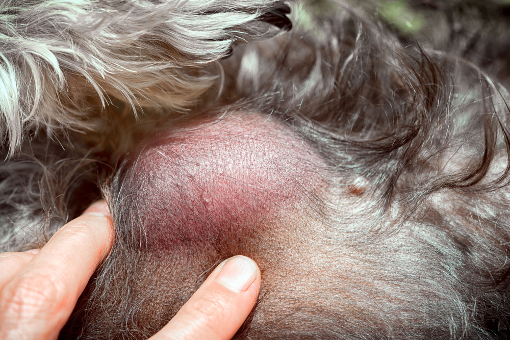
1. Non-Cancerous Lumps
- Abscess: An abscess is a pocket of pus. An infection under the skin can cause an abscess, usually secondary to a bite or injury. Grass awns tunneling through the skin can also cause an abscess.
- Hematoma: A hematoma is a blood clot that can occur under the skin after an injury or surgery. Another common example is an aural hematoma, which can happen when a dog has an ear infection. Their constant head shaking leads to the rupture of blood vessels in the ear, causing the hematoma.
- Inflammation: A lump can be made of inflammatory cells. An insect bite can sometimes lead to inflammation that looks like a lump.
- Seroma: A seroma is a build-up of clear fluid under the skin and occurs sometimes after surgery.
2. Benign Lumps
- Histiocytoma: A histiocytoma is a benign growth that can go away on its own. It is common in younger dogs. It is often raised, hairless, and sometimes ulcerated.
- Lipoma: A lipoma is an abnormal growth of fatty tissue that doesn’t spread but can get quite large. They are sometimes removed to prevent them from bothering the dog.
- Apocrine Gland Cysts: These cysts originate from the glands in your dog’s skin. They most commonly occur on the head and neck, and dogs sometimes have multiple cysts.
- Melanocytoma: A darkly pigmented benign skin lump that is typically small. They are most commonly seen on the head and forelimbs of middle-aged or older dogs. Melanocytomas arise from melanin-producing cells.
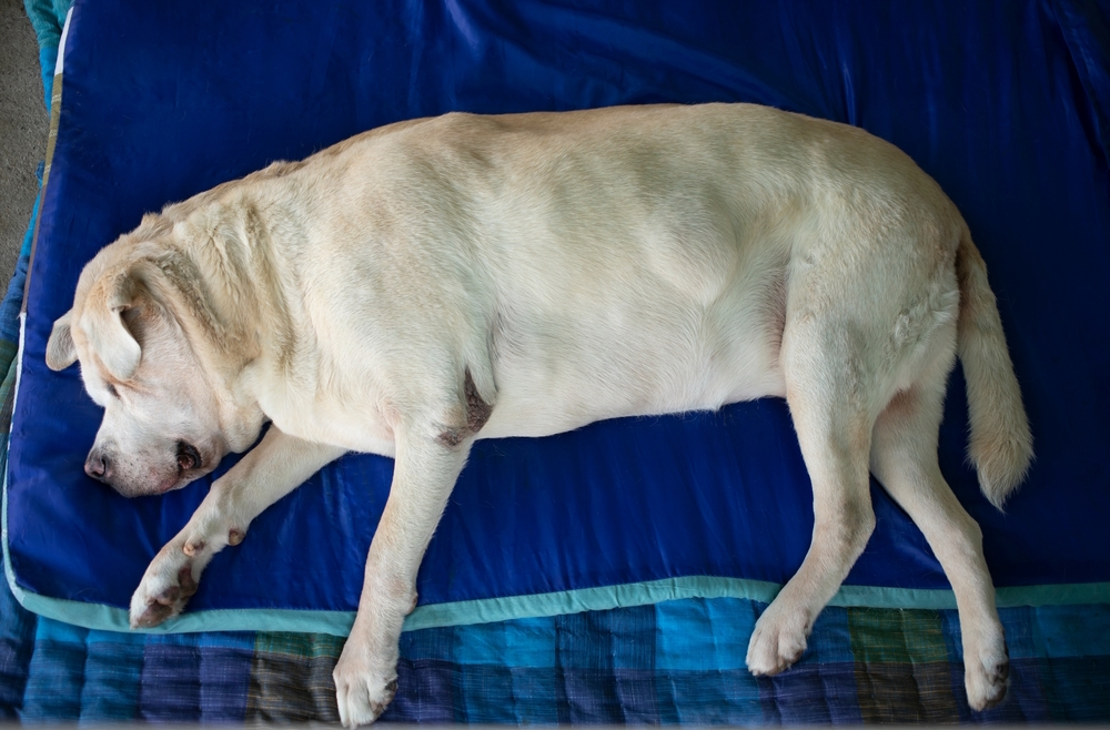
3. Cancerous Lumps
- Mast Cell Tumors: Tumors arising from mast cells in the skin are graded from I to III, with III being the most malignant form. These cancers can release histamine, leading to signs similar to allergic reactions. They don’t have a “standard” appearance and can look like a lipoma or histiocytoma.
- Soft Tissue Sarcomas: These cancers arise from connective tissue and infiltrate the healthy tissues around them. They can sometimes spread to other areas of the body.
- Malignant Melanoma: This is a malignant form of melanocytoma. It usually occurs around the lips, mouth, and nail beds but can appear anywhere in the body. Despite its origins in pigment-producing cells, it isn’t always pigmented. It is very invasive and metastasizes.
If you’re concerned about your pet’s well-being, we recommend you contact a veterinarian.
PangoVet. It’s an online service where you can <b>talk to a vet online</b> and get the personalized advice you need for your pet — all at an affordable price!</p>
<p><div class="su-button-center"><a href=https://www.dogster.com/ask-the-vet/"https://pangovet.com/?utm_source=dogster&utm_medium=article&utm_campaign=dog_infection_illness%22 class="su-button su-button-style-default" style="color:#FFFFFF;background-color:#FF6600;border-color:#cc5200;border-radius:9px;-moz-border-radius:9px;-webkit-border-radius:9px" target="_blank" rel="nofollow"><span style="color:#FFFFFF;padding:0px 24px;font-size:18px;line-height:36px;border-color:#ff944d;border-radius:9px;-moz-border-radius:9px;-webkit-border-radius:9px;text-shadow:none;-moz-text-shadow:none;-webkit-text-shadow:none"> Click to Speak With a Vet</span></a></div></div></div></p>"}" data-sheets-userformat="{"2":513,"3":{"1":0},"12":0}"> If you need to speak with a vet but can’t get to one, head over to PangoVet. It’s an online service where you can talk to a vet online and get the personalized advice you need for your pet — all at an affordable price!
What to Do if Your Dog Has a Lump
If your dog has a lump, check its size and note the location. You will need to take your dog to the vet to get the lump checked, but it’s helpful to give the vet an accurate history. Your vet will want to know how long the lump has been there and if it is growing.
If you think the lump is growing, try to estimate how much it has grown. For example, has it doubled in size, or has the size increased by a certain percentage? If the lump is bleeding or looks ulcerated, when did this start? If you have noticed any signs of illness in your dog, like coughing or lethargy, you should let the vet know.
Large neoplasias don’t appear suddenly, so if you have noticed a golf ball-sized lump on your dog overnight, it’s possible that the lump has been there longer than you think or the lump is caused by something other than neoplasia.
You should do a full body check on your pet for further lumps. Start at their head, running your fingers through their fur, and work your way over their body, legs, and tail. You should do this regularly to look for new lumps, especially in older dogs. Finding a cancerous lump early can save your dog’s life.

What to Expect at the Vet
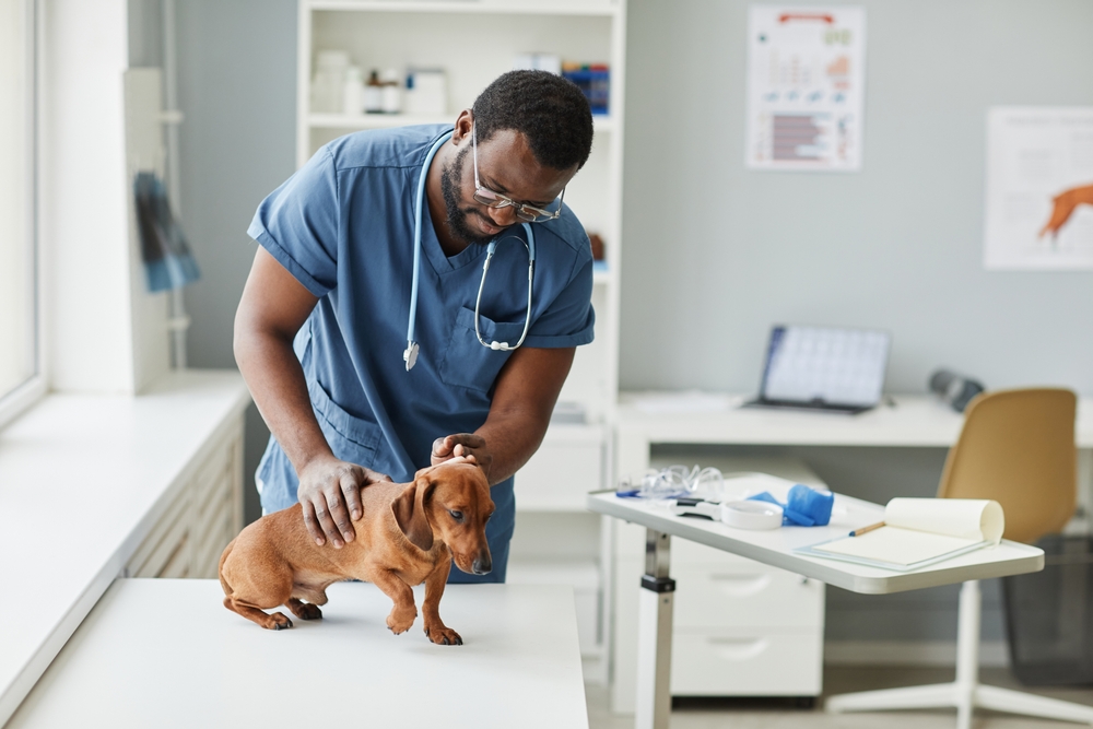
Your vet will examine your dog and the lump. Most vets will take a sample using a fine needle aspirate. This means puncturing the lump with a needle and sucking out some of the cells to examine under a microscope. Sometimes, they can simply press the lump onto a microscope slide to get a sample, but it only works if it oozes.
These samples may give a diagnosis, but it is often difficult to say 100% as, despite being able to see the cells, you can’t see how they are organized in the tissue. For example, it can be hard to differentiate between a malignant melanoma and a melanocytoma just by looking at the individual cells since they arise from the same cell type.
Your vet may recommend monitoring the lump or surgery to remove or biopsy the lump. An excisional biopsy involves the vet trying to take out the whole lump and sending it away for testing at a veterinary lab. Veterinary pathologists will diagnose the lump.
When a cancerous lump is removed, the goal is to remove a portion of healthy tissue around the lump that is tumor-free; this is known as the “margin.” Microscopically, cancer cells can extend centimeters away from the visible lump, so clean margins are a good sign of complete lump removal.
An incisional biopsy is sometimes performed when the lump is large or difficult to remove. A small portion of the lump is sampled and sent away for testing. If monitoring the lump is the best option, check it weekly.
To keep track of it, measure its diameter with a ruler. If the lump changes, communicate any changes to your vet. Some dogs get so many lumps that it is hard to keep track of them, so we recommend keeping a log of them somewhere if you aren’t removing them.
Conclusion
Skin lumps and bumps are common, and many are nothing to worry about. Unfortunately, some skin lumps can be cancerous, and you can’t tell just by looking whether a lump falls into this category. All lumps should be vet-checked because removing a cancerous lump early can save your dog’s life. You should also keep track of your dog’s lumps and bumps so you can be informed of any changes.
Featured Image Credit: KingTa, Shutterstock


